
Medical imaging has always played a crucial role in diagnosing and treating various health conditions. Over the years, advancements in medical technology have revolutionized the way doctors and healthcare providers detect and monitor diseases. One of the most recent breakthroughs in this field is Manhiascan, an innovative medical imaging technology that offers precision, speed, and safety in diagnosing a wide range of conditions.
In this article, we will explore Manhiascan in detail, examining how it works, its applications, benefits, and its potential to reshape the future of medical diagnostics.
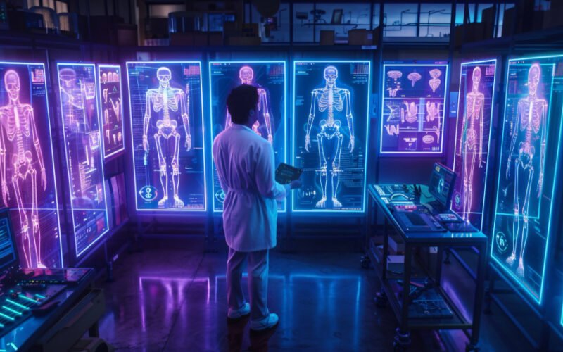
What is Manhiascan?
Manhiascan is an advanced form of medical imaging technology that combines elements of magnetic resonance imaging (MRI), computed tomography (CT), and ultrasound to create highly detailed, 3D images of the body’s internal structures. This technology is designed to offer higher resolution images with more accurate data while minimizing the exposure of patients to radiation.
Unlike conventional imaging technologies that might rely on one type of scan, Manhiascan integrates multiple imaging methods to provide comprehensive diagnostic insights. It is particularly useful in the early detection of diseases and in monitoring the progression of existing medical conditions, making it a valuable tool for healthcare providers.
How Does Manhiascan Work?
Manhiascan works by combining several different imaging techniques into a single, unified scan. This process begins with the use of magnetic fields and radio waves, similar to what is used in MRI scans, to create high-resolution images of soft tissues. These images are further enhanced by incorporating data from CT scans, which use X-rays to visualize hard tissues such as bones. Finally, ultrasound technology is used to provide real-time imaging of blood flow and organ movements.
The integration of these different technologies allows Manhiascan to create a complete, three-dimensional image of the targeted area. This unified image is much more detailed and accurate than what can be achieved through any single imaging method, offering medical professionals a clearer picture of the patient’s condition.
The scan process itself is non-invasive, and in many cases, it can be completed in less time than traditional imaging methods. Patients undergoing a Manhiascan experience less discomfort, and because the technology reduces the need for repeated scans, it is considered safer in the long term.
Applications of Manhiascan in Healthcare
Manhiascan is versatile and can be used across various medical fields. Below are some of the key areas where this technology is currently being applied:
1. Oncology (Cancer Detection and Treatment)
Manhiascan is particularly useful in the early detection of cancer. By providing high-resolution images of tumors, it allows oncologists to detect even the smallest growths that might be missed by other imaging methods. Furthermore, Manhiascan can be used to monitor the progression of cancer during treatment, helping doctors adjust therapies based on real-time data.
This technology is especially effective in imaging soft tissues, such as the lungs, liver, and brain, where tumors can often go unnoticed in the early stages. By catching these tumors early, patients have a better chance of receiving treatment that can prevent the cancer from spreading.
2. Neurology (Brain and Nervous System Disorders)
Neurological disorders such as Alzheimer’s disease, Parkinson’s disease, and multiple sclerosis are notoriously difficult to diagnose and monitor. Manhiascan’s ability to produce highly detailed images of the brain and nervous system makes it an invaluable tool for neurologists. It can detect subtle changes in brain structure and function, helping doctors diagnose neurological conditions early and track their progression over time.
The real-time imaging provided by Manhiascan also helps in surgical planning for conditions like brain tumors, ensuring that surgeons have precise data on the location and size of the affected area.
3. Cardiology (Heart and Blood Vessel Imaging)
In the field of cardiology, Manhiascan is used to image the heart and blood vessels. It can detect conditions like blockages, aneurysms, and heart defects with a high degree of accuracy. By integrating ultrasound technology, it also provides real-time data on blood flow and heart function, which can be critical for diagnosing conditions such as congestive heart failure or coronary artery disease.
The detailed 3D images allow cardiologists to understand the structure and function of the heart in ways that were not possible before. This technology can also guide minimally invasive procedures like angioplasty and stenting, reducing the need for open-heart surgery.
4. Orthopedics (Bone and Joint Imaging)
Manhiascan is beneficial for imaging bones and joints, making it ideal for use in orthopedics. Conditions like fractures, arthritis, and sports injuries can be visualized with incredible clarity, allowing doctors to accurately assess the extent of the damage and plan appropriate treatments.
Unlike traditional X-rays, which might miss subtle bone changes, Manhiascan can detect microfractures and early signs of osteoporosis, ensuring that patients receive treatment before the condition worsens.
5. Gastroenterology (Digestive System Imaging)
Manhiascan’s ability to image soft tissues makes it useful in the field of gastroenterology as well. Doctors can use it to visualize organs such as the stomach, intestines, liver, and pancreas, detecting conditions like ulcers, inflammation, and tumors. The integration of real-time imaging allows for better monitoring of conditions like Crohn’s disease and irritable bowel syndrome (IBS).
Also, explore Mansrufer: Male Grooming With Innovative Techniques
Advantages of Manhiascan Over Traditional Imaging
This medical imaging technology offers several key advantages over traditional imaging technologies:
1. Higher Image Resolution
Manhiascan provides much higher image resolution compared to standard imaging methods. This increased resolution allows for more accurate diagnoses, as even the smallest abnormalities can be detected.
2. Comprehensive Imaging
By integrating multiple imaging technologies, Manhiascan offers a comprehensive view of the body. Rather than relying on separate scans for different areas or conditions, doctors can obtain a complete picture in one session.
3. Reduced Radiation Exposure
One of the biggest concerns with traditional CT scans is the exposure to radiation. Manhiascan minimizes this risk by combining imaging techniques that use less radiation. This makes it a safer option, especially for patients who require frequent scans, such as those undergoing cancer treatment.
4. Faster Diagnosis
It reduces the time it takes to get results. The technology is designed to produce images quickly, allowing doctors to make faster diagnoses and start treatment sooner. This can be especially important in emergencies, such as stroke or heart attack, where every minute counts.
5. Non-Invasive and Painless
The entire scanning process is non-invasive and painless, providing comfort for patients. Manhiascan does not require incisions, injections, or the use of contrast dyes, which are sometimes needed in traditional imaging procedures.
Challenges and Future Potential of Manhiascan
While Manhiascan is a promising advancement in medical imaging, there are still challenges to overcome. One of the primary concerns is the cost of the technology. The equipment required for Manhiascan is expensive, and this cost may be passed on to patients, making it less accessible to everyone. Additionally, as with any new technology, there is a learning curve for medical professionals who need to be trained in its use.
Despite these challenges, the potential for Manhiascan is immense. As the technology becomes more widespread, costs are likely to decrease, making it more accessible to hospitals and clinics around the world. The ability to provide fast, accurate, and comprehensive imaging could revolutionize how medical conditions are diagnosed and treated, leading to better patient outcomes.
Conclusion
Manhiascan represents the future of medical imaging technology. By integrating multiple imaging methods into one system, it offers superior resolution, faster diagnosis, and improved patient safety. Its applications across fields like oncology, neurology, cardiology, and orthopedics demonstrate its versatility and effectiveness in diagnosing a wide range of conditions.
As this technology continues to evolve, it is expected to become a cornerstone of modern medicine, offering healthcare professionals the tools they need to provide accurate and timely diagnoses. For patients, this means better care, earlier detection of diseases, and more effective treatment plans, ultimately improving their quality of life.